TMJ/TMD
-Temporomandibular
Disorders and Orofacial Pain
Diagnosis and
Treatment
What is TMJ or
TMD and what are its symptoms?
The temporomandibular (TMJ) jaw joints in front of the
ear are the hinges between the jawbone and head. Neck movement, breathing,
talking, chewing, swallowing, smiling, all require jaw movement. As the lower
jaw moves, it requires both TMJ's to move and slide freely. When there is an
abnormality with the joints and associated muscles of the jaw, pain can
develope. Symptoms of TMJ Disorders include the following:
- Clicking, popping joints/frank noise
- Headache, neck pain, face pain
- Chew pain, swallowing difficulty
- Ear Pain, blocked eustachian tubes
- Pain behind eye
- Locking, inability to open, dislocation
- Dizziness, tinnitus/ear ringing
- Muscle soreness in temple region, side of face, neck
- Awaking with sore clenched jaws
- Throat pain
Headache and orofacial pain are often complex,
involving both local areas and other regions of the musculoskeletal system of
the body. Thus, a thorough history and
comprehensive clinical examination are done at the
first visit. At chairside, clinical evaluation and charting involves the
structures inside the mouth and outside. Palpation
of the Temporomandibular joints, muscles of mastication, and entire head/ neck
region is critical in assessing orofacial/TMJ related pain and headache.
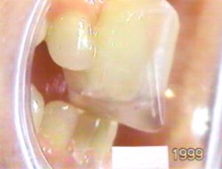
Range of Motion of the jaw in all dimensions is
measured, correlated to stethescopic sounds. The movement of opening and
closing should be smooth without jarring or deflection. Lateral side to side
and forward motion should be unstrained. When there is restriction and
locking/popping of the joint, the existing closed position of the mandible and
the fit of the teeth are manually compared to other potential jaw positions.
This is done to test for differences in TMJ disk/joint function im-provement.
Normal opening of 50 mm is seen here, without deviation to the side or joint
noise.
Diagnostic procedures will be recommended such as
impressions of the teeth/jaws, from which dental study models are made.
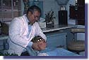
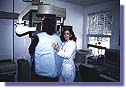
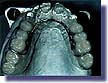
Radiographic/X-Ray imaging is done to view jaw
structures and abnormalities of the teeth,TMJ, head and neck region. A
panoramic radiograph of the jaw is seen being taken
here:
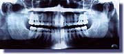
Panoramic Radiograph shows entire jaw-healthy
structures-normal view seen in this photo:
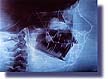
A complete orthodontic/orthopedic facial skeletal
analysis with anatomic tracings is used to help correlate teeth positions and
jaw posture to boney growth. This is seen from the lateral skull,
cephalometric x-ray view:
Besides palpation assessment, other procedures may
involve measurement of muscle dysfunction and contraction/spasm via an EMG, or
electromyogram, to the head, neck and jaw region. Tightness produces
contraction and muscle referred pain.After a thorough diagnostic workup, and
case assessment/plan discussion, a conservative course of treatment is
usually recommended. This may typically involve an initial pharmacologic
approach to acute pain and physical medicine
modalities, such as moistheat/ hydrocollator pack , cold/vapocoolant
spray & stretch, EGS electrogalvanic stimulation, TENS transcutaneous
electrical stimulation and ultrasound.
The use of light wave monochromatic Infrared energy
for wound healing has been well established over the past 40 years. Both
visible and infrared light have been shown to effect at least 24 different
positive changes at the cellular level. With absorption of therapeutic light
waves, unique therapeutic effects in tissue occur via photobiochemical
reactions. Indeed vision, photosynthesis, tanning, and vitamin D metabolism are
responses to wave-length dependent light reactions. Increase of vascularity
with new capillary formation, stimulation of collagen production for damaged
tissue repair, release of ADP for cellular metabolism/energy, increase of
lymphatic system activity, with increase of lymphatic vessel size and flow for
edema/swelling reduction. Increases RNA and DNA synthesis, reduction of
excitability of nerve tissue, pain relief, stimulation of fibroblastic activity
to aid repair, increased phagocytosis in blood cell "cleanup"/ scavenging
activity, critical in infection control. Thermal effect, without cellular heat
from the diodes; stimulation of granulation and connective tissue for ulcer and
wound healing, with acetylcholine release and parasympathetic effects.
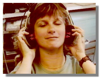
There has been recent development for Orofacial
region/TMJ pain , with new infrared treatment modality delivery. The
applicability to the jaw/TMJ region and cervical is done through two hand
pieces mounted inside the medical device, which looks like stereo headphones;
this houses the multiple light emitting phototherapy/low energy laser diodes.
Treatment of joints and deep muscle is approached directly, with each side is
treated as a unit. With the headphone-like dual assembly infrared system, there
is delivery of bilateral side-to-side therapy.
Manipulation/mobilization procedures to jaw and neck
region, trigger point injections, home care exercises and postural
stress/tension reduction are also utilized to help with long-range
stabilization. These are often integrated with an initial in-traoral daytime
orthosis/ soft mouthguard and then a nightguard.
Splint Appliances: A hard
laboratory processed splint is usually prescribed for longer term use, 24 hours
a day in acute situations. The goal is to reduce pressure on the joint to get
it to settle down (decompression).
**** Further more detailed
information available through patient/consumer information
catalogue****
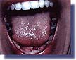
A Full arch maxillary splint
appliance is seen here, with functional overlay occlusion/bite for
chewing. Biting surfaces help guide jaw downward and forward to reduce and
reposture disk of TMJ. Anterior coverage is clear for daytime esthetics. Same
principles of design for mandibular splint apply. For night time only, a "pull
forward ramp" may be added, with breathing holes. Arch bar and clasping provide
for additional retention; a flat-planed design for primary muscle/clenching
type pain may be elected. Return visits require further bite/appliance
adjustments, coupled with therapy. Long-range plans may call for other dental
rehabilitation, orthodontic, and prosthodontic considerations.
Referral may be made for an MRI or magnetic resonance image of the joint, where
potential surgery is anticipated for dislocation or chronic displacement.
Maintenance/return visits with continued therapy and splint/niteguard use help
prevent relapses.
NTI tm
DEVICE
(Nociceptive Trigeminal
Inhibition)
Clenching Suppression System
A very successful new night appliance is the
“NTI TM” clenching suppression system. Patients with aching jaws,
morning headache, sore, sensitive teeth, neck stiffness, TMJ pain, and tension
headache often unknowingly clench their teeth at night. The NTI device prevents
the temporalis and masseter muscles from forcefully contracting utilizing the
jaw opening reflex. This allows the appliance to protect incisors, posterior
molars, surrounding tissues and TM joints, and helps prevent associated
headaches of muscle contraction. The system and its patents have been FDA
approved for headache and TMJ therapy, and is a recommended procedure for
migraine head pain treatment by many international headache centers/clinics
using a medical/dental team approach.
The appliance design is to reduce muscle
contraction headache, with excessive masticatory myalgia and muscle
hypertonicity relaxation, focusing on the masseters and temporalis. The
appliance helps reduce the net effect of clenching and tooth grinding stress
and wear on the teeth and occlusion. Contact is anterior at the incisor region,
with the cuspid, bicuspid, and molars out of contact upon closure. The incisal
region features a smoothe functional sliding ramp to accommodate multiple jaw
positions at night.
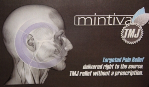
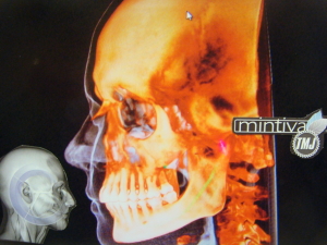
Mintiva
TMJ
Targeted pain relief delivered right to the source
Mintiva TMJ is a new topical treatment for TMJ pain,
featuring a proprietary deliver system that absorbs quickly, effectively
relieves pain, and allows greater range of motion of the jaw. TMintiva is an
over -the- counter topical cream, developed to dental professionals'
specifications to target and relieve temporomandibular joint pain. It is
non-greasy, fast acting, and does not leave a medicinal smell like some topical
treatments.Other Mintiva formulations can also relieve arthritic neck and back
joint pain. The cream is applied to the TMJ, jaw muscles, massaged into the
skin. For further information and ordering, click on the following link, using
coupon discount code #1003: www.mintiva.com.